If you are citizen of an European Union member nation, you may not use this service unless you are at least 16 years old.
You already know Dokkio is an AI-powered assistant to organize & manage your digital files & messages. Very soon, Dokkio will support Outlook as well as One Drive. Check it out today!
• observe the similarities and differences between bacterial, protist, animal, and plant cells.
• recognize the different shapes and characteristics of prokaryotic cells.
• recognize the typical structures in animal cells.
• recognize the typical structures in plant cells.
• learn to recognize and use the parts of a compound microscope.
• determine magnification power.
• focus a microscope.
• make a wet mount.
B. Textbook Correlation:
Please review Chapter 4: General Features of Cells when preparing for the lab.
C. Introduction to observing cells using a microscope:
Cells were first discovered in 1665, when an English biologist, Robert Hooke, was studying cork under a primitive compound microscope he had made. He actually observed cell walls because cork cells are dead and have lost their internal components. Hooke coined the word cell, derived from the Latin word cellula, meaning small compartment, to describe the structures he observed. The cell theory was later developed in the mid-1800s when a German botanist named Matthias Schleiden was studying a plant under a microscope and observed that it had many similar looking compartments. Later, when discussing this with his friend Schwann, a physiologist, Schwann recalled possibly also seeing cell-like structures in animals. After research, Schleiden’s hypothesis was expanded to involve plants and animals.The cell theory, or cell doctrine, which is credited to both Schleiden and Schwann with contributions from Virchow, has three parts. 1. All living organisms are composed of one or more cells. 2. Cells are the smallest units of life. and 3. New cells come only from pre-existing cells by cell division.
There are two basic types of cells, prokaryotes and eukaryotes. Prokaryotes have a rather simple structure. They a membrane bound nucleus. Nearly all species have a cell wall. Inside the cell wall is a plasma membrane. Inside the membrane is the cytoplasm, nucleoid region, and ribosomes. Eukaryotic cells, on the other hand have a more complex structure. They tend to be larger and have membrane bound organelles. This gives the cells compartmentalization and organization. Cells are classified into six main kingdoms of life which are: Archaebacteria, Eubacteria, Protista, Fungi, Plantae, and Animalia. Organisms are placed into these categories based on similarities or common characteristics. Some of the characteristics that are used to determine placement are cell type, metabolism, and reproduction. Archaebacteria evolved 3-4 billion years ago are unicellular and are prokaryotes. They are found in extreme environments such as places with no oxygen or places that are highly acidic. Eubacteria also evolved 3-4 billion years ago are unicellular and prokaryotes. They are very common in our day to day lives. Protista, Fungi, Plantae, and Animalia are all Eukaryotes. Protista evolved 1.5 billion years ago and are unicellular. Protists are the cells that mainly to not belong to any of the other categories. Fungi evolved 1 billion years ago. They can be multicellular or unicellular. Some examples include mushrooms, mold, and mildew. Plantae evolved 500 million years ago. They are multicellular and are found in plants. Animalia evolved 700 million years ago are multicellular and are found in animals.
However most cells are so small they cannot be seen with the naked eye, instead, they must be viewed through a microscope.The microscope is a magnification tool that enables researchers to study the structure and function of cells. The microscope was first invented in 1595 by Zacharias Jansen of Holland. Ever since it has been being refined and modified to create a better clearer image. In 1509 Galileo,father of modern physics and astronomy, heard of these early experiments, worked out the principles of lenses, and made a much better instrument with a focusing device. In 1675 the Dutch merchant Anton van Leeuwenhoek refined techniques of making lenses and was able to observe single-celled microorganisms such as bacteria.A major advance in microscopy occurred in 1931 when Max Knoll and Ernst Ruska invented the first electron microscope. During the early 20th century both microscopes improved dramatically, advancing our understanding of cells.
There are two main types of microscopes, light microscopes and electron microscopes. A light microscope utilizes light for illumination whereas an electron microscope utilizes electrons for illumination. Electron microscopes have a far better resolution but are also much more expensive and therefore not as commonly used in classroom settings.
This picture shows the structure of a typical prokaryotic cell. Notice that it lacks internal compartmentalization. The main similarities between the two types of cells include the plasma membrane, cytoplasm, and ribosomes.
This picture shows two different kinds of eukaryotic cells, an animal cell and a plant cell. The main difference between these two cells in the cell wall and the chloroplasts in the plant cell. Notice that both cells are much more complex then the prokaryote cell and that they both have many membrane bound organelles.
Study the following list when preparing to use a microscope:
1. Carry a microscope by supporting it in both hands in an upright position. One hand should grasp the microscope arm while the other provides a flat platform for its base. Place it down on a flat surface that has been cleared of debris.
2. If your microscope has a rheostat, which adjusts light intensity, make sure it is turned to minimum light intensity before turning on the light.
3. Clean all lenses by wiping with grit-free lens paper. Never use a paper towel or any other material to clean the lens to avoid making permanent scratches.
4. When you adjust focus, always begin with the lowest power first. Avoid contacting the lens to the microscope slide on the stage.
5. If your microscope is not working as you expect, notify your lab instructor immediately. Do not attempt to make repairs by removing or adjusting parts.
The Basic Parts and Functions of the Compound Microscope
As you examine the compound microscope, review Table 1, in which each part is listed and described. In the last column of the table, indicate which number on the microscope diagram corresponds to the location of that particular part.
Magnification in the Microscope
With two-lens systems, there is a simple method to figure the total magnification when using each objective. The total magnification is equal to the magnification of the ocular multiplied by the magnification of the objective.
Resolution.
Resolution refers to the ability to distinguish two points as separate points. As you might think, resolution is dependent upon the distance between the two points. When using a microscope or any optical instrument, the closer the points are, the more difficult it becomes to see them as separate points. Numerical aperture is related to resolution, although magnification has very little to do with it. The higher the numerical aperture, the better the resolution will be. An object can be magnified larger and still be blurry, because although magnification has increased the resolution has not improved.
Focusing the Microscope:
There are some basic steps to follow when focusing the microscope.
1. Always begin with the objective that has the lowest magnification—the scanning objective. Turn the revolving nosepiece until this objective is over the stage.
2. Use the coarse adjustment knob to move the stage so that it is closest to the lens.
3. Place a slide on the stage and secure it with the clips.
4. While looking through the ocular, slowly move the stage away from the lens using the coarse adjustment knob.
5. The diaphragm can be used to increase or decrease the amount of light coming through the stage opening.
6. Be sure that you have the object clearly focused at this point.
7. Now you are ready to move to the next objective. The term parfocal is used to describe a microscope’s ability to remain in focus no matter what objective is used. The key to this parfocal ability is to always begin with the scanning objective.
8. Keep in mind that as you move up in magnification, you will not be able to use the coarse adjustment knob. The objectives are too long and will crack the slide. Use only the FINE ADJUSTMENT KNOB.
9. Rotate the revolving nosepiece to the next objective—low power. Use only the fine adjustment knob to sharpen the focus.
10. Now that you have the slide in focus at this power, move the revolving nosepiece to the next objective—high power—and repeat the process described in step 9.
11. The oil immersion lens can only be used with immersion oil. It is commonly needed to see very small objects such as bacteria.
Your First Observation: Observing Prokaryotic Cells
Prokaryotes have existed for at least 3.8 billion years and are thought to be the first cells on earth. Bacteria are the most common prokaryotes. There are more bacteria on and within our bodies than we have human cells. Most bacteria are non-pathogenic (non-disease causing) and actually are used in making a variety of useful products. Bacteria can take a variety of shapes and sizes. Three basic shapes commonly found among members of this kingdom include bacillus, coccus, and spiral. The term bacillus (bacilli plural) describes a bacterium that is rod shaped or cylindrical. An example of a bacillus shaped bacterium includes Clostridium botulinum, the causative agent of botulism and the source of the drug Botox. A bacterium that is symmetrically spherical is termed coccus. Examples of cocci shaped bacteria include Streptococcus pyogenes, the causative agent of strep throat, and Staphylococcus pneumoniae, the causative agent of both bacterial pneumonia and meningitis. Spiral shaped bacteria have one or more twists. A spirochete is a bacterium with multiple twists that gives it a corkscrew-like appearance. Treponema pallidum is a spirochete that causes the STD syphilis.
Bacteria can also take a variety of arrangements. Recall that these are single celled organisms. They reproduce through cell division. In some cases, after the cells divide, they remain in contact with one another. For example, the arrangement of two cells that remain in contact is termed diplo-. Bacteria arranged as long chains are called strepto- and bacteria that are clustered are described as staphylo-. For example, if you observe bacillus shaped cells arranged in a chain, you would describe them as steptobacillus.
Beside shape and arrangement, bacteria are commonly classified by their cell wall structure. Some bacteria have a thickened cell wall structure made of the carbohydrate peptidoglycan. This type of cell wall is referred to as a Gram (+) cell wall. Other bacteria have a very thin layer of peptidoglycan that is surrounded by an additional outer membrane. These bacteria are called Gram (-) bacteria. The origin of these names comes from a staining technique developed by Hans Christian Gram. In this technique, bacteria with the thickened cell wall are colored purple and the bacteria with the outer membrane are colored red/pink. This method is used extensively as a diagnostic tool to identify bacteria.
Procedure:
1. Observe the slides that demonstrate the three different shapes of bacteria. Sketch the shapes and arrangements you observe in the space provided below.
Coccus
Bacillus
Spirillum
2. Observe the Staphylococcus aureus specimen and draw in the space provided. Describe the shape, arrangement, and cell wall of this bacterium in the space provided.
Whole View: Bacteria is arranged in colonies.
Each colony can contain over 1 million.
One Colony. Still one big yellow blur.
Practicing Your Craft: Observing Yeast
It is time to practice using the microscope. We will use a member of the Fungi kingdom called Saccharomyces cerevisiae in this exercise. Members of the Fungi kingdom are eukaryotic and can be unicellular (yeast) or multicellular (molds). Their cell walls contain the polysaccharide chitin. They play the role of decomposers in the ecosystem and are essential for recycling nutrients back to the soil. In this exercise, we will observe the yeast S. cerevisiae using three different objective lenses.
1. Using the steps for focusing, examine the specimen using the scanning objective. How do they look? Draw what you view below.
The specimen looks like a web of cells, arranged in circular patterns, with a open white space in the middle of each "circle" of cells
2. Now view the specimen using the low power objective. How do they look?
Stained nuclie is visible, and the cells are arranged in small random groups. Lots of negative space.
3. Finally, examine the cells using the high power objective. How do they look?
Yeast is clearly visible in the specimen.
4. Explain the differences that you have seen using each objective.
S. cervisiae (Scanning Objective)
Total Magnification = 40X
S. cervisiae (Scanning Objective)
Total Magnification = 100X
S. cervisiae (Scanning Objective)
Total Magnification = 400X
In this picture, it can bee seen that the cells are arranged in a net type arrangement. Individual cells were not visible. Each visible "strand" is made or multiple "strands". There is a lot of negative space.
The "strands" are arranged in a random pattern. The cells are arranged in clusters. It is similar to the 40X magnification
In this picture, the cells are clearly visible. They are arranged in clusters and appear circular in shape. The way the cells are arranged, forms a lot of negative space. Each individual cells is clearly prominent. In many cells, the nucleus and the vacuole can be identified.
Preparing A Wet Mount
Most of the microscope slides that you will be studying in the lab will be prepared slides. That is, they have been sectioned, dehydrated, and stained by professional histologists and purchased for your educational use. Staining brings out the structural details of specimen.
In some cases, a wet mount slide may be prepared in order to view objects that do not require sectioning and dehydration for observation. The advantage is its simplicity and quickness, providing a sample that can be observed within minutes of obtaining it. Clinical labs use this procedure with body fluids, such as blood and urine. In this exercise, you will learn how to make a wet mount slide.
Protist cells:
Many protists inhabit fresh water and are characterized by their mode of movement. The three basic motile structures found in protists include flagella, cilia, and pseudopods. A few are pathogenic like Plasmodium vivax, the causative agent of malaria and Giardia intestinalis, the causative agent of giardiasis. Most are free living and will be the focus of this part of the lab.
Follow the instructions below to prepare and observe a wet mount:b
3. Many of the microorganisms that live in fresh water are characterized by their motility. Start with the scanning objective to provide the greatest field of view. After finding a specimen, observe at low power and draw what you find. Describe the mode of movement observed.
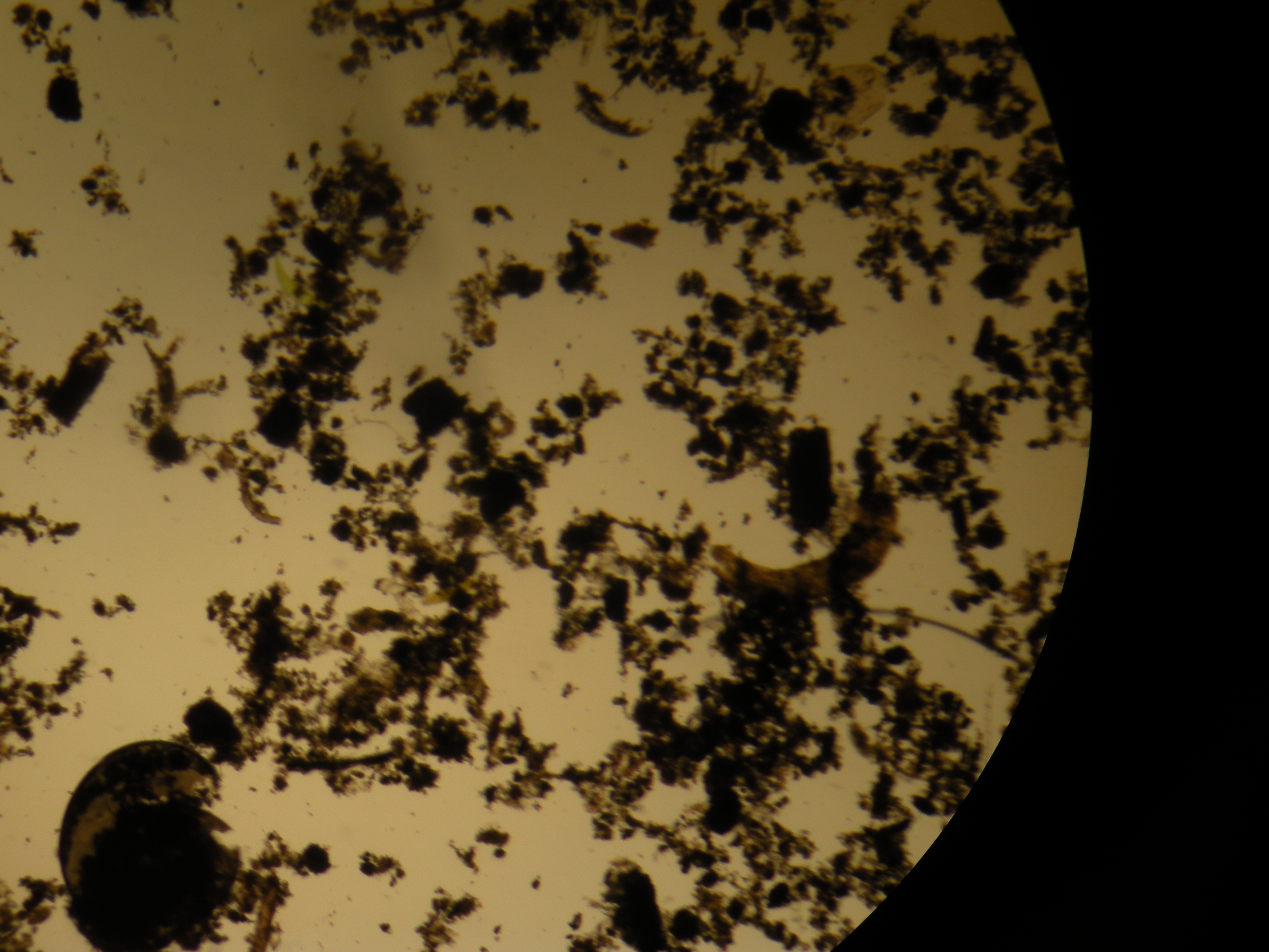
Pond Water 40X
100X: A long worm like specimen was slowly moving through the medium
400X: A amoeba moving very fast through the water, mostly staying by the algae
Plant cells:
1. Use your tweezers to remove a small leaf from the growing tip of an Elodea sprig.
2. Make a wet mount.
3. Locate the cells with low power. Then focus on a small number of cells using high power.
4. Locate the following structures and label them in your sketch below:
• chloroplasts: small bright green organelles
• cell wall
• central vacuole: this will appear as a clear area without chloroplasts
• nucleus: it is slightly larger than the chloroplasts and is clear or very light brown – NOT green
This is a live video of the plant cells under the microscope. The moving particles are Chloroplasts.
5.Also observe cytoplasmic streaming in these cells as indicated by chloroplasts moving about the cell, usually around the outer edge of the cell.
6. To better view some of the cell structure, place a drop of methylene blue stain at the edge of the coverslip. Then touch a piece of tissue to the opposite side of the coverslip to draw the stain under the coverslip.
7. Reexamine the leaf cells for a better defined cell wall.
8. Next, cut an onion bulb into quarters and then separate the layers.
9. Take one layer and peel away the very thin inner membrane.
10. Make a wet mount with this thin membrane. You may need to use the tweezers to spread out the membrane without wrinkles.
11. Place the slide on your microscope and use low power to locate the cells. Then switch to high power to observe a few cells more closely.
Animal Cells:
1. Using the broad end of a toothpick, lightly scrape the inside of your cheek.
2. Stir the scraping in a drop of methylene blue on a microscope slide. Cover with a coverslip.
3. Place the slide on the microscope stage and find the cells using low power. Then switch to high power.
4. Identify the nucleus, cytoplasm, and plasma membrane. Remember, you can’t actually see the plasma membrane but can identify its location as the outer boundary of the cell.
5. Sketch a typical cell below and label nucleus, cytoplasm,and plasma membrane.
The picture above is of stained cheek cells. The nucleus is clearly visible and the border is the plasma membrane.
Question:
List similarities and differences between plant and animal cells that you observed today.
Similarities
a. Nucleus
b. Plasma membrane
Differences
a. No cell wall in animal cells
b. No chloroplast in animal cell
c. No large vacuole in animal cell
This exercise was adapted from Brooker, R. Biology (2nd edition). McGraw-Hill Publishing, New York.
A. Learning objectives:
Students will assess the relationship between symptoms presented by patients and specific cellular functions (Evaluation).
Students will infer malfunctioning cellular organelles as being the cause of illness (Analysis).
Students will explain how the malfunctioning of particular organelles can result in a given array of symptoms (Analysis).
Students will interpret data and assign functions to a family of aquaporins (Evaluation and Analysis).
B. Pre-lab Assignment: Read through the introduction to this exercise and become familiar with the assignment.
C. During Lab: A group of four will discuss the four cases, with each member taking turns as the facilitator. After completing the case, the group will present their findings on their researched disease. There will be plenty of time to accomplish this goal during the 160 minutes provided for lab.
D. Post-Lab Assignment: Upload the PowerPoint presentation to the site using SlideShare for others to view for future reference and complete the assigned questions in the case Study.
E. Organelles and Illness:Eukaryotic cells are characterized by membrane-bound organelles that compartmentalize the cytoplasm. This physical segregation of areas within the cytoplasm allows eukaryotic cells the ability to perform several functions, some that are biochemically contrary to one another, simultaneously—increasing the overall biochemical efficiency of the cell. Most of the processes conducted by organelles are accomplished through complex metabolic pathways involving multiple enzymes, rather than single-step chemical processes. Typically, an overall cellular activity is the result of a coordinated effort of many organelles.
Improper functioning of organelles can be the cause of diseaseand illness. Tay-Sachs disease, for example, is a fatal neurological disease caused by malfunctioning lysosomes. Lysosomes are organelles that serve to break down complex materials (like proteins, lipids, nucleic acids and carbohydrates) either that are taken up from outside the cell or are the product of chemical reactions within the cell. The resultant molecules are then utilized by the cell as molecular building blocks, or are eliminated. In Tay-Sachs disease a genetic mutation causes a deficiency or malfunction in the production of one of the enzymes needed to break down a class of lipids found in brain cells. As the lipids accumulate in brain cells, neurological function is impaired. Symptoms typically begin to show around three to six months after birth and include irritability, seizures, blindness, deafness and paralysis. Most affected individuals die by the age of five. The disease is currently incurable, but experimental gene therapy is currently an area that is being explored as a possible mechanism for treatment.
As the overall functions conducted by organelles are typically the result of complex metabolic pathways, diagnosing disorders associated with particular organelles can be difficult. Because cellular organelles conduct multiple activities, the symptoms associated with their malfunction are diverse and often impact multiple body systems. Plants (all eukaryotes in fact) can suffer from disorders due to malfunctioning organelles.
Imagine that you are part of a research team that specializes in diagnosing disorders associated with malfunctioning cellular organelles and structures. Medical doctors consult your group to help diagnose particularly difficult cases. In the next class meeting you will work in small collaborative groups of four to provide a cellular explanation for several particularly difficult cases. Each person in the group will act as the ‘facilitator’ for one case, leading the group discussion, promoting input from each of the other students (who will be acting as ‘discussants’) and formalizing the group response. In the role of a discussant, students provide their knowledge, experience and perspectives, compare and contrast the inputs of other members of the group and collaborate in the formulation of the group response. Once the source of the problem has been identified, research for specific diseases caused by defects in the specific organelle. In the example above, a defective lysosome in nerve cells results in Tay-Sachs disease. At the end of the activity, you will be called on to present your group’s answers to one of the cases (not necessarily the one you were the facilitator for) and give a 5-10 minute presentation on the disease that you researched. You will have time in lab to complete both tasks You will act as both a facilitator and a discussant in the activity.
Case Study #1: _______________________________ (Facilitator)
Rationale for Diagnosis (explanation of how history, physical examination and laboratory results suggest diagnosis):
Case Study #2:_______________________________ (Facilitator)
2A. Patient History:
55 year-old female
Excellent health to date
Psychological stress
Recently won lottery
Marital stress
Recent onset of:
Fatigue
Headaches
Excessive salivation
Nausea
Vomiting
Diarrhea
Vertigo
Abdominal pain
Weight loss
2B. Physical Examination
Swelling of eyelids
Partial limb paralysis
Hyperkeratosis (increased pigmentation) of skin on palms & soles
White banding on fingernails
Muscle weakness
2C. Laboratory Results
low levels of ATP production
Unusually high levels of arsenic
Abnormal levels of cell death—not due to apoptosis (programmed cell death)—rather cell death appears to be due to lack of energy to maintain homeostasis due to low ATP levels.
Well the dissecting microscope we were using only went up to 400x.
Were we suppose to use a prepared slide and a light microscope? If we were, we completely missed it!
You don't have permission to comment on this page.


Comments (4)
Derek Weber said
at 11:02 pm on Nov 3, 2010
Explain the differences in yeast at different magnifications.
Pankhuri Garg said
at 11:32 pm on Nov 3, 2010
Edited :)
Derek Weber said
at 4:36 am on Dec 11, 2010
What happened to the picture of S. aureus at 1000x total. You have only the colony picture here.
Pankhuri Garg said
at 8:15 am on Dec 11, 2010
Well the dissecting microscope we were using only went up to 400x.
Were we suppose to use a prepared slide and a light microscope? If we were, we completely missed it!
You don't have permission to comment on this page.